Vocal Health, Wellness & Understanding
Now, maybe you don’t want to hear this or…maybe you already know…Exercising is just as important for voices as for the rest of our bodies. When we want to increase our physical abilities we simply have to exercise. The same goes for our singing voices. When we warm up our voices before singing, we avoid the dangers of straining our voices or damaging our vocal chords.
Our vocal chords are just two strips of soft tissue, located within the larynx. This area together with our breath is responsible for all our vocal sound, so it’s important that we develop and care for it properly. Just as we need to stretch and relax the muscles before we run or lift weights, we need to warm up our voices. Warming up loosens those crucial muscles, reduces the risk of injury and can sometimes even help to remove excess mucous! (Eeeew! Better out than in!) ;-)
Vocal exercises will help us look after your precious singing voices whilst giving you increased technical vocal abilities.
The Vocal Chords
from Wikipedia
The vocal folds, also known commonly as vocal cords or voice reeds, are composed of twin infoldings of mucous membranestretched horizontally, from back to front, across the larynx. They vibrate, modulating the flow of air being expelled from the lungs during phonation.[1][2][3]
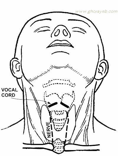
Open during inhalation, closed when holding one’s breath, and vibrating for speech or singing (oscillating 440 times per second when singing A above middle C), the folds are controlled via the vagus nerve.
Structure
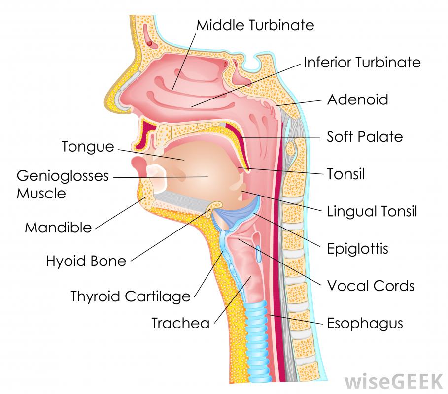
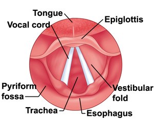
The vocal folds are located within the larynx at the top of the trachea. They are attached posteriorly to the arytenoid cartilages, and anteriorly to the thyroid cartilage. They are part of the glottis which includes the rima glottidis. Their outer edges are attached to muscle in the larynx while their inner edges, or margins are free, forming the opening called the rima glottidis. They are constructed from epithelium, but they have a few muscle fibres in them, namely the vocalis muscle which tightens the front part of the ligament near to the thyroid cartilage. They are flat triangular bands and are pearly white in color. Above both sides of the glottis are the two vestibular folds or false vocal folds which have a small sac between them.
Situated above the larynx, the epiglottis acts as a flap which closes off the trachea during the act of swallowing to direct food into the esophagus. If food or liquid does enter the trachea and contacts the vocal folds it causes a cough reflex to expel the matter in order to prevent pulmonary aspiration.
Variation
Males and females have different vocal fold sizes. Adult male voices are usually lower pitched due to longer and thicker folds. The male vocal folds are between 1.75 cm and 2.5 cm (approx 0.75″ to 1.0″) in length,[2] while female vocal folds are between 1.25 cm and 1.75 cm (approx 0.5″ to 0.75″) in length. The vocal cords of children are much shorter than those of adult males and females. The difference in vocal fold length and thickness between males and females causes a difference in vocal pitch. Additionally, genetic factors cause variations between members of the same sex, with males’ and females’ voices being categorized into voice types.
In newborns
Newborns have a uniform monolayered lamina propria, which appears loose with no vocal ligament.[14] The monolayered lamina propria is composed of ground substances such as hyaluronic acid and fibronectin, fibroblasts, elastic fibers, and collagenous fibers. While the fibrous components are sparse, making the lamina propria structure loose, the hyaluronic acid (HA) content is high.
HA is a bulky, negatively charged glycosaminoglycan, whose strong affinity with water procures HA its viscoelastic and shock absorbing properties essential to vocal biomechanics.[15] Viscosity and elasticity are critical to voice production. Chan, Gray and Titze, quantified the effect of HA on both the viscosity and the elasticity of vocal folds (VF) by comparing the properties of tissues with and without HA.[16] The results showed that removal of HA decreased the stiffness of VF by an average of 35%, but increased their dynamic viscosity by an average of 70% at frequencies higher than 1 Hz. Newborns have been shown to cry an average of 6.7 hours per day during the first 3 months, with a sustained pitch of 400–600 Hz, and a mean duration per day of 2 hours.[17] Similar treatment on adult VF would quickly result in edema, and subsequently aphonia. Schweinfurth and al. presented the hypothesis that high hyaluronic acid content and distribution in newborn VF is directly associated with newborn crying endurance.[17] These differences in newborn vocal fold composition would also be responsible for newborns inability to articulate sounds, besides the fact that their lamina propria is a uniform structure with no vocal ligament. The layered structure necessary for phonation will start to develop during the infancy and until the adolescence.[14]
The fibroblasts in the newborn Reinke’s space are immature, showing an oval shape, and a large nucleus-cytoplasm ratio.[14] The rough endoplasmic reticulum and Golgi apparatus, as shown by electron micrographs, are not well developed, indicating that the cells are in a resting phase. The collagenous and reticular fibers in the newborn VF are fewer than in the adult one, adding to the immaturity of the vocal fold tissue.
In the infant, many fibrous components were seen to extend from the macula flava towards the Reinke’s space. Fibronectin is very abundant in the Reinke’s space of newborn and infant. Fibronectin is a glycoprotein that is believed to act as a template for the oriented deposition of the collagen fibers, stabilizing the collagen fibrils. Fibronectin also acts as a skeleton for the elastic tissue formation.[14] Reticular and collagenous fibers were seen to run along the edges of the VF throughout the entire lamina propria.[14] Fibronectin in the Reinke’s space appeared to guide those fibers and orient the fibril deposition. The elastic fibers remained sparse and immature during infancy, mostly made of microfibrils. The fibroblasts in the infant Reinke’s space were still sparse but spindle-shaped. Their rough endoplasmic reticulum and Golgi apparatus were still not well developed, indicating that despite the change in shape, the fibroblasts still remained mostly in a resting phase. Few newly released materials were seen adjacent to the fibroblasts. The ground substance content in the infant Reinke’s space seemed to decrease over time, as the fibrous component content increased, thus slowly changing the vocal fold structure.
These are vocal chords…I promise!
Watch the vocal chords up close as this quartet sings a hymn in four-part harmony
In adults
Human VF are paired structures located in the larynx, just above the trachea, which vibrate and are brought in contact during phonation. The human VF are roughly 12 – 24 mm in length, and 3–5 mm thick.[18] Histologically, the human VF are a laminated structure composed of five different layers. The vocalis muscle, main body of the VF, is covered by the mucosa, which consists of the epithelium and the lamina propria.[19] The latter is a pliable layer of connective tissue subdivided into three layers: the superficial layer (SLP), the intermediate layer (ILP), and the deep layer (DLP).[5] Layer distinction is either made looking at differential in cell content or extracellular matrix (ECM) content. The most common way being to look at the ECM content. The SLP has fewer elastic and collagenous fibers than the two other layers, and thus is looser and more pliable. The ILP is mostly composed of elastic fibers, while the DLP has fewer elastic fibers, and more collagenous fibers.[19] In those two layers, which form what is known as the vocalis ligament, the elastic and collagenous fibers are densely packed as bundles that run almost parallel to the edge of the vocal fold.[19]
The extracellular matrix of the VF LP is composed of fibrous proteins such as collagen and elastin, and interstitial molecules such as HA, a non-sulfated glycosaminoglycan.[5]While the SLP is rather poor in elastic and collagenous fibers, the ILP and DLP are mostly composed of it, with the concentration of elastic fibers decreasing and the concentration of collagenous fibers increasing as the vocalis muscle is approached.[19] Fibrous proteins and interstitial molecules play different roles within the ECM. While collagen (mostly type I) provides strength and structural support to the tissue, which are useful to withstanding stress and resisting deformation when subjected to a force, elastin fibers bring elasticity to the tissue, allowing it to return to its original shape after deformation.[5] Interstitial proteins, such as HA, plays important biological and mechanical roles in the VF tissue.[15] In the VF tissue, HA plays a role of shear-thinner, affecting the tissue viscosity, space-filler, shock absorber, as well as wound healing and cell migration promoter. The distribution of those proteins and interstitial molecules has been proven to be affected by both age and gender, and is maintained by the fibroblasts.[5][9][15][20]


